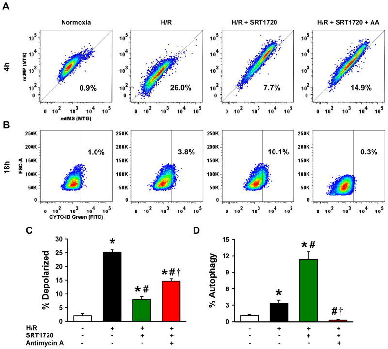
Figure 9.
SRT1720-induced autophagy is dependent on mitochondrial membrane potential after hypoxia-reoxygenation (H/R) in H4IIE hepatocytes. H4IIE hepatocytes underwent 6 h of oxyrase-induced hypoxia in glucose-, pyruvate- and serum-free media followed by 4 h or 18 h of reoxygenation in complete media. At the beginning of reoxygenation, cells were treated with 0.125% DMSO (vehicle), 500 nM of SRT1720, or 500nM of SRT1720 plus 500 nM of antimycin A. (A, C) After 4 h of reoxygenation, cells were stained with mitotracker green and mitotracker red FM dye for measurement of mitochondrial mass (mtMS) per cell and mitochondrial membrane potential (mtMP) per cell, respectively. Cells below the reference line (thin diagonal line) were considered as cells with depolarized mitochondria. (B, D) After 18 h of reoxygenation, cells were stained with CYTO-ID green autophagy dye, arbitrary gate drawn at 1% baseline autophagy rate in the normoxia control cells (vertical thin line). Data are presented as means ± SE (n = 6/group), representative of 3 independent experiments. * P < 0.05 versus control; # P < 0.05 versus H/R, † P < 0.05 versus H/R plus 100 nM SRT1720. FSC-A, forward scatter - area.

