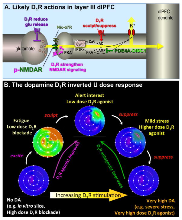
Figure 1. Schematic illustration of DA D1R influences on Delay cell firing in layer III of the primate dlPFC.
A. Localization of D1R in layer III dlPFC pyramidal cell networks. Pyramidal cells interconnect via NMDAR synapses on spines, with permissive actions from nicotinic α7 receptors (nic- α7R). Immunoelectron microscopy has shown that D1R are concentrated on dendritic spines, where they can be seen directly within the synapse (magenta), and near the synapse where they often co-localize with HCN or KCNQ potassium channels (red). The open state of both of these channels is increased by cAMP-PKA signaling. Physiological recordings from monkeys indicate that D1R activates feedforward cAMP-PKA-calcium signaling, which opens K+ channels and weakens nearby synaptic inputs (red). At optimal doses this sculpts away noise from irrelevant inputs, but at higher doses, e.g. as occurs during stress, it causes nonspecific suppression of Delay cell firing and loss of working memory. Feedforward cAMP-calcium signaling is held in check by the phosphodiesterase, PDE4A, which is anchored in place by DISC1 (Disrupted In Schizophrenia). Studies in nonprimate species suggest that D1R within the synapse phosphorylate NMDAR via activation of cAMP-PKA and PKC signaling; this maintains NMDAR in the synaptic membrane and strengthens connections (magenta). There are also D1R on glutamate axon terminals that may reduce glutamate release (purple). For detailed description, see Arnsten et al, 2015. The asterisk indicates the spine apparatus, the extension of the smooth endoplasmic reticulum into the spine.
B. A schematic illustration of the DA D1R inverted U influence on the “memory fields” of dlPFC Delay cells. For details, see (5). Under optimal arousal conditions, Delay cells generate persistent representations of visual space, displaying high rates of firing (orange-red) to the memory of one spatial location, and low rates of firing (blue) to the memory of all other spatial locations. When there is no D1R stimulation, Delay cells have little firing. Low levels of D1R stimulation appear to be excitatory, e.g. phosphorylating NMDAR to increase their trafficking into the synapse (7). This can produce noisy firing for all directions, as represented by the generalized green-orange coloring of the memory field. With optimal levels of D1R stimulation, there are additional sculpting actions, gating out “noise”, e.g. by opening a subset of HCN channels. At still higher levels of D1R stimulation (e.g. as occurs during stress), there is excessive HCN channel opening and Delay cell firing is generally suppressed. Under these conditions the neuron is not able to generate persistent representations of visual space.

