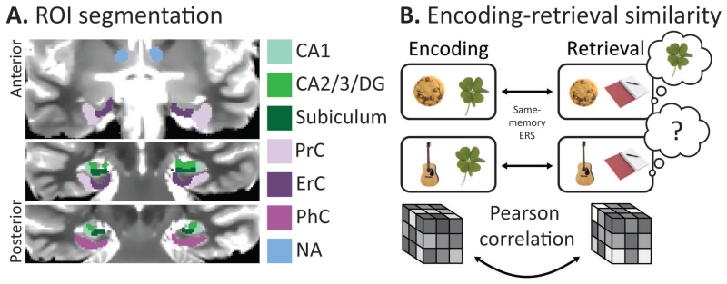
Figure 2. fMRI methods.
A. An example of the ROI boundaries over a T2 weighted scan. All ROIs were hand-drawn while referencing coronal slices over T1 and T2 weighted images. B. A schematic of the ERS analysis. We computed same-memory ERS for each trial by correlating its pattern of activation at encoding and retrieval.

