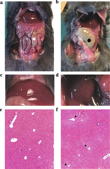
Fig. 7.

Aspect of peritoneal cavity of the sham-operated mice (a, c, e) and the 20 % ligated mice (b, d, f) 24 h after surgery. a The cecum is identified by a white star. b The 20 % ligated animal with cecal abscess ( ). When observing liver macroscopic morphology, we found a patchy appearance corresponding to pale ischemic areas (d) in contrast to the sham liver (c). f These areas were centrilobular necrosis of hepatocytes (asterisk) (e and f hematoxylin-eosin coloration ×50)
