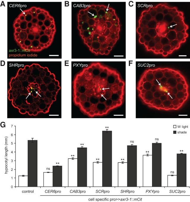
Figure 2.

axr3-1 expression in different cell types alters hypocotyl growth in shade. (A–F) Representative fluorescence images of hypocotyl cross-sections of 9-d-old seedlings expressing axr3-1::mCit protein (green) under the control of different cell-specific promoters and counterstained with propidium iodide (red). CER6pro was used to drive axr3-1 expression in the epidermis (A), CAB3pro in green tissue (including strong cortical and weak epidermal expression) (B), SCRpro in the endodermis (C), SHRpro throughout the stele (D), PXYpro in procambium cells (E), and SUC2pro in the phloem companion cells (F). Arrows mark examples of nuclear-localized axr3-1::mCit. Not all nuclei of a given cell layer are in the plane of focus. In our hands, PXYpro had an expression pattern similar to that of SHRpro, including expression in the meristematic pericycle and procambium and perhaps other unidentified cells of the vasculature. SUC2pro also had occasional weak expression in the epidermis. For each promoter, the cell-specific pattern of axr3-1::mCit expression was unaffected by shade, and multiple independent lines had similar phenotypes that correlated with expression level (Supplemental Figs. S2, S3). In addition to hypocotyl expression, these promoters drove axr3-1::mCit in equivalent cell types of the cotyledons and, in some cases, various zones of the root. Bars, 50 µm. (G) Hypocotyl length in white (W) light and shade of 9-d-old seedlings expressing axr3-1::mCit under the control of a cell-specific promoter. Control is hemizygous for UAS::axr3-1::mCit. Data show mean ± SEM. Means are compared with the control in the same light condition using Tukey's HSD. (**) P < 0.005; (ns) not significant.
