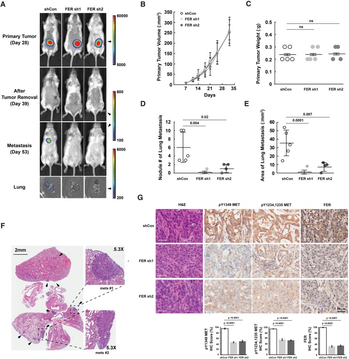
Figure 6.
Loss of FER reduced lung metastasis burden of ovarian cancer cell xenografts with inactivation of MET. (A) At the indicated time points, mice injected with CAOV4 cells expressing either shCon (n = 5), FER sh1 (n = 5), or FER sh2 (n = 4) were imaged using IVIS bioluminescence imaging. Representative images are shown. (B,C) Measurements of subcutaneous tumor volume (B) and weight (C) for mice injected with either shCon, FER sh1, or FER sh2 CAOV4 cells. (D) Metastasis lesion count (naked eye as well as IVIS imaging confirmation) with mice injected with either shCon, FER sh1, or FER sh2 CAOV4 cells. (E) Metastasis lesion area measurement with mice injected with either shCon, FER sh1, or FER sh2 CAOV4 cells. (F) Representative H&E staining of lung sections from mice with metastatic lesions that express the control shRNA (shCon). (G) H&E and immunohistochemistry staining (pTyr MET and FER) of subcutaneous tumor sections from mice injected with CAOV4 cells expressing either shCon, FER sh1, or FER sh2. The immunohistochemistry image was scored with Aperio software.

