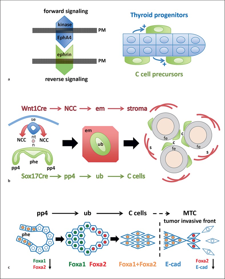
Fig. 2.
C cell development in mouse thyroid. a The interaction of C cell precursors with follicular progenitor cells influences the final number of thyroid C cells (based on data published by Andersson et al. [23]). After integration of the ultimobranchial bodies with the embryonic thyroid only follicular cells express EphA4, a tyrosine kinase receptor that binds promiscuously to ephrin-type ligands on adjacent cells. Genetic deletion or kinase inactivation of EphA4 leads to reduced C cell numbers postnatally. Whether EphA4 on follicular cells affects C cells directly (by altered reverse signalling of ephrins) or indirectly (e.g. by a paracrine growth factor) is unknown. b Lineage tracing proves thyroid C cells originate in endoderm (Johansson et al. [1]). By recombination of a reporter mouse with Cre lines that distinguish endoderm (Sox17Cre) and neural crest (Wnt1Cre) lineages, respectively, it is evident that C cells derived from Sox17+ anterior endoderm, specifically from the pharyngeal pouches from which the paired ultimobranchial bodies develop. In contrast, the neural crest gives rise to ectomesenchymal cells that surround the ultimobranchial body and later, after fusion with embryonic thyroid, converts to the stromal compartment of the gland (see also paper by Kameda et al. [14]). c Differential expression of forkhead box transcription factors in different C cell lineage stages (Johansson et al. [1]). Foxa1 and Foxa2, required in concert for proper endoderm formation foregoing organogenesis, are co-expressed in the pharyngeal endoderm except in the pouch domains from which the ultimobranchial bodies develop. Lineage tracing reveals that all pouch progenitors originate in endoderm proper indicating Foxa1 and Foxa2 are specifically down-regulated as the ultimobranchial bodies form and delaminate. The free ultimobranchial bodies re-express Foxa1 and Foxa2 in a distinct pattern that reveals Foxa1+ (Foxa2-) proliferating and Foxa2+ (Foxa1-) non-proliferating domains of the ultimobranchial epithelium. Eventually, all ultimobranchial cells and embryonic C cells co-express Foxa1 and Foxa2. Invasive cells of human medullary thyroid carcinoma transiently lose Foxa1 and Foxa2 expression. Loss of Foxa2 characterizes epithelial-to-mesenchymal transition accompanied by down-regulation of E-cadherin (E-cad), a key feature of the invasive tumour phenotype. This differs malignant from normal C cells and C cell precursors that express E-cadherin in all developmental stages. a-c PM = Plasma membrane; se = surface ectoderm; nt = neural tube; n = notochord; NCC = neural crest cells; phe = pharyngeal endoderm; pp4 = fourth pharyngeal pouch; em = ectomesenchyme; ub = ultimobranchial body; c = C cells; fe = follicular epithelium; s = stromal cells.

