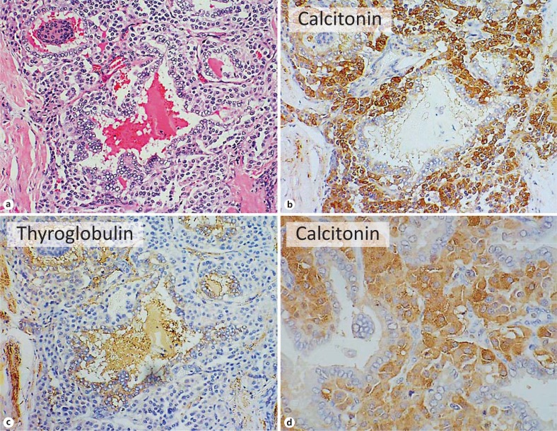
Fig. 3.
Mixed medullary-papillary carcinoma of the thyroid. a-c Near serial sections. a HE. b Immunohistochemistry for calcitonin. c Immunohistochemistry for thyroglobulin. d High-power view of calcitonin-positive cells. Note irregular follicles with papillary projections lined by epithelium and separated by sheets of neoplastic cells. The lining epithelial cells show the nuclear features of papillary carcinoma and both lining cells and luminal colloid are positive for thyroglobulin, while the cells between and spreading around the follicular structures are positive for calcitonin.

