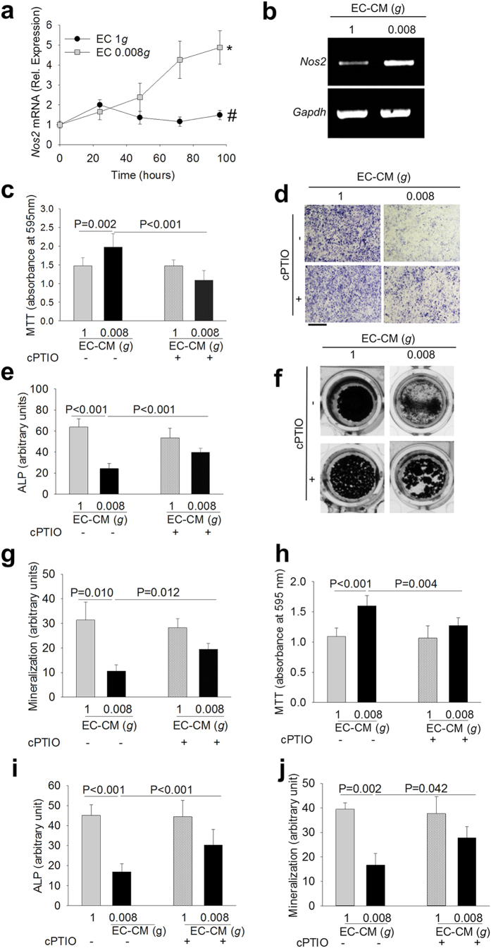
Figure 3. Role of the NOS2-NO-COX2 pathway in endothelial cell-osteoblast crosstalk.
Mouse primary endothelial cells were subjected to 1g and 0.008g for the indicated times. (a) Real time RT-PCR analysis of Nos2 normalized versus Gapdh. (b) EA.hy976 cells were subjected to 1g and 0.008g for 96 hours. RT-PCR of Nos2 normalized versus Gapdh. Mouse primary osteoblasts were treated with 1g- or 0.008g-EC-CM in the absence or presence of the NO quencher cPTIO. (c) MTT proliferation assay. (d) Representative images of ALP cytochemical staining. (e) Densitometric quantification of ALP staining. (f) Representative images of mineralized nodule formation by the Von Kossa staining. (g) Densitometric quantification of mineralization. Frozen 1g- and 0.008g-EC-CM were de-frozen in ice and used to treat mouse primary osteoblasts in the absence or presence of the NO quencher, cPTIO. (h) MTT proliferation assay. (i) Densitometric analysis of ALP cytochemical staining. (j) Densitometric quantification of mineralization (Von Kossa staining). Data are the mean ± SD of 3 independent experiments (Student’s t test). Bar = 100 μm. All gels have been run under the same experimental conditions.

