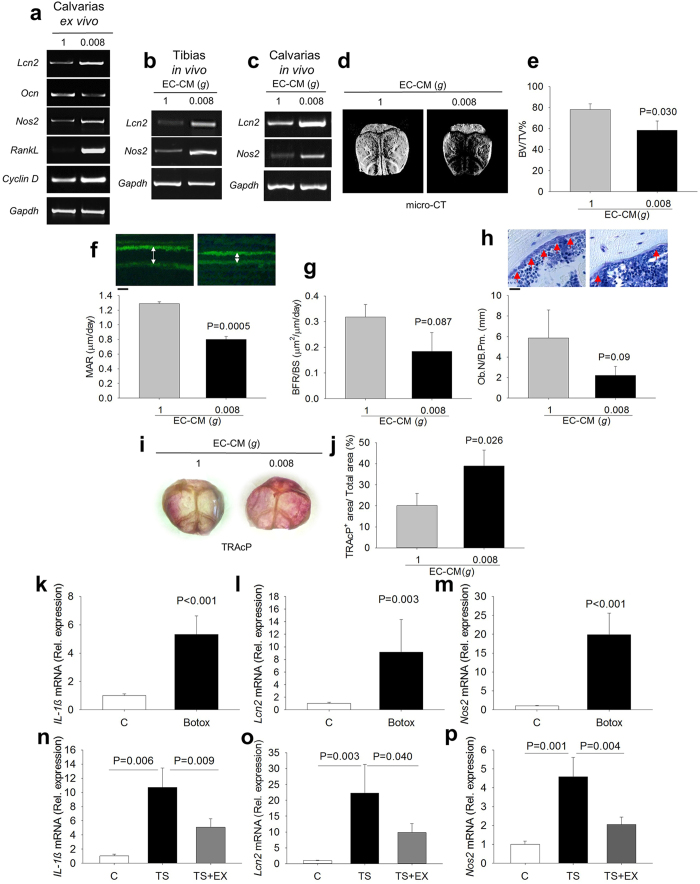
Figure 7. Ex vivo and in vivo activation of the IL-1β/NOS2/LCN2 pathway.
(a) Calvarias were isolated from 7-day old C57BL/6J mice and cultured ex vivo with 1g- or 0.008g-EC-CM. RT-PCR of Lcn2, Ocn, RankL, Nos2, and Cyclin D1 normalized versus Gapdh. 1g- and 0.008- EC-CM were injected in 4 week-old C57BL/6J mice (b) into tibias (intra-tibial injection) and (c) onto calvarias (subcutaneous injection). RT-PCR of Nos2 and Lcn2 normalized versus Gapdh. (d) Representative of micro-CT images of 4 week-old C57BL/6J mouse calvarias treated in vivo with 1g- and 0.008g-EC-CM. (e) Quantitative analysis of calvarial bone volume over total tissue volume (BV/TV). (f) Calcein labeling (green fluorescence, double white arrows) (upper panels) and quantification of Mineral Apposition Rate (MAR) (lower panel). (g) Quantification of Bone Formation Rate (BFR). (h) Histological images of toluidine blue stained histological sections (osteoblasts, red arrows) (upper panels) and quantification of osteoblast number over bone perimeter (Ob.N./B.Pm.) (lower panel), (i) Representative images of whole mount TRAcP staining and (j) TRAcP quantification. Eight-week old C57BL/6J mice were subjected to Botox injection as described in methods, RNA was isolated from their long bones and mRNA expression of (k) IL-1β, (l) Lcn2 and (m) Nos2 was analyzed by real-time RT-PCR. Eight-week old C57BL/6J mice were subjected to hind limb tail suspension (TS), with or without physical exercise (TS + EX) by the forced swim test to counteract the mechanical unloading. Total RNA was isolated from femurs and subjected to real time RT-PCR for (n) IL-1β, (o) Lcn2 and (p) Nos2. Images are representative and data are the mean ± SD of at least 3 independent experiments or 3 mice/group (Student’s t test). Bar = (f) 2 μm; (h) 20 μm. All gels have been run under the same experimental conditions.

