Figure 5.
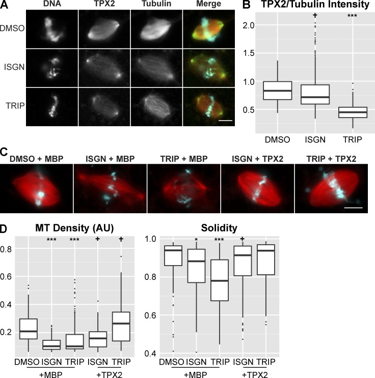
Addition of TPX2 partially rescues spindle defects caused by perturbing ncRNA biogenesis. (A) Example images of TPX2 staining in control and inhibitor-treated reactions. In the merged image, microtubules are red, TPX2 is green, and DNA is cyan. Bar, 10 µm. (B) Plot showing the ratio of TPX2/tubulin intensity in spindles in control (n = 242) and inhibitor-treated reactions (ISGN n = 234, TRIP n = 120). The TPX2/tubulin ratio decreased 13.9% in ISGN-treated extract and 45.9% in TRIP-treated extract. (C) Example images of spindles assembled in extract after treatment with DMSO + 200 nM MBP (n = 193), ISGN + 200 nM MBP (n = 235), TRIP + 200 nM MBP (n = 199), ISGN + 200 nM TPX2 (n = 265), or TRIP + 200 nM TPX2 (n = 126). Bar, 10 µm. (D) Quantification of spindle microtubule density and spindle solidity under each of the conditions in C. Median microtubule density decreased 50.6% in MBP + ISGN–treated extract, 51.0% in MBP + TRIP–treated extract, 24.0% in TPX2 + ISGN–treated extract, and increased 26.9% in TPX2 + TRIP–treated extract. +, P < 0.05; *, P < 10−5; **, P < 10−10; ***, P < 10−15 (Kolmogorov–Smirnov test).

