Figure 5.
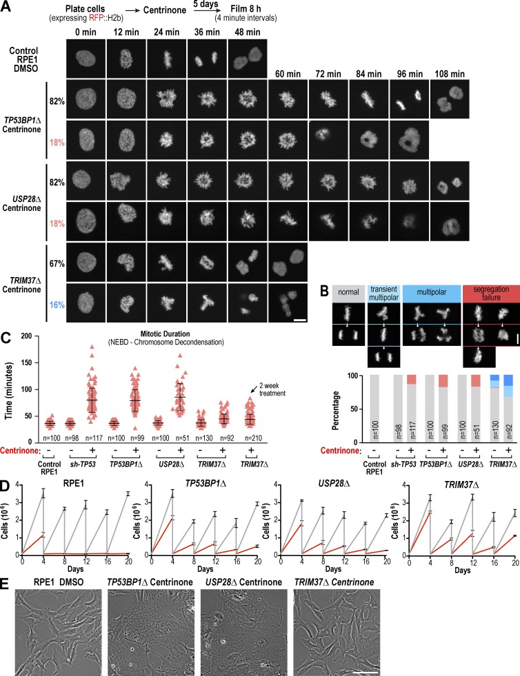
Knockout of TRIM37, but not TP53BP1 or USP28, suppresses mitotic defects in centrosomeless cells. (A) Selected images from timelapse series of representative DMSO-treated control RPE1 cells and centrinone-treated TP53BP1Δ, USP28Δ, and TRIM37Δ mutant cells, acquired as outlined in the schematic. (B) Graph plotting the distribution of mitotic phenotypes observed for each condition along with representative images. (C) Graph plotting mitotic duration. Individual cell values (red triangles) are shown along with the mean and SD (black bars) for each condition. NEBD, nuclear envelope breakdown. (D) Graphs plotting the results of passaging assays monitoring the growth of control and knockout RPE1 cell lines after acute addition at day 0 of DMSO (vehicle) or centrinone. (E) Representative phase-contrast images of fields of DMSO-treated control RPE1 and knockout mutant cells after prolonged (>20 d) treatment with centrinone. Bars: (A and B) 10 µm; (E) 100 µm.

