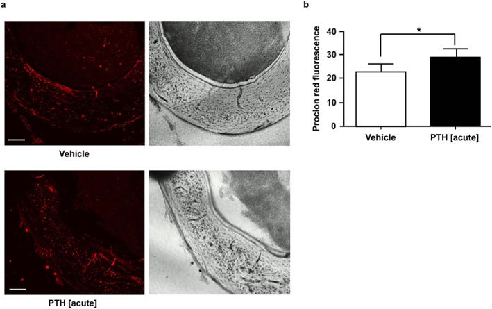
Figure 2.

Acute effect of PTH treatment on lacunar–canalicular perfusion. (a) Representative confocal micrographs of non‐decalcified sections of the femoral mid‐diaphysis showing the uptake of fluorescent tracer in cortical bone 5 min after injection of vehicle (control) and PTH 1–34 (80 µg/kg). Scale bar = 100 µm. (b) Average tracer fluorescence within a region of interest manually drawn within the postero‐medial quadrant of the mid‐diaphysis cortical bone. Values are mean ± SD, n = 4 mice/group; * P < 0.05 versus Control
