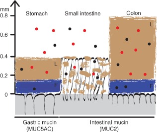
Figure 2.

Mucus organization and nanoparticle interactions in the gastrointestinal tract. The gastrointestinal tissue is represented in grey with black folds representing the structure interfacing the lumen. The predominant mucin isotype expressed in each region is shown in parenthesis. L denotes the loosely bound outer mucus layer. Non‐interacting lamellar strands of loosely bound mucus in the small intestine are also shown in brown. F denotes the firmly attached inner mucus layer, shown in blue. Mucoadhesive nanoparticles are represented by the red circles; non‐mucoadhesive nanoparticles are represented by the black circles. (Reprinted with permission from Ref 22. Copyright 2011 National Academy of Sciences)
