
Figure 2.
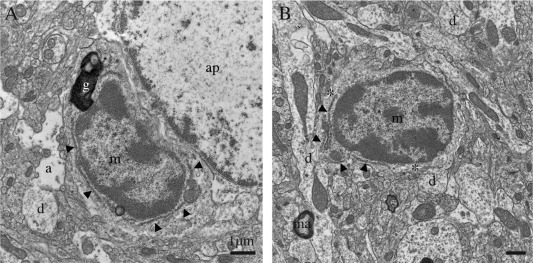
Examples of normal microglia, observed in the median eminence of the hypothalamus in a nontransgenic control mouse. A, B: Unstained microglia (m) generally display a lighter cytoplasm and nucleoplasm with a clearly defined chromatin pattern, compared with the dark microglial cells. They also share with the dark microglia a small elongated nucleus delineated by a narrow nuclear cistern, associated pockets of extracellular space (asterisks), distinctive long stretches of endoplasmic reticulum (arrowheads), frequent endosomes, lipofuscin granules (g), and cellular inclusions. a = astrocytic process, ap = astrocytic perikaryon, d = dendrites, and ma = myelinated axon. Scale bars = 1 μm.