
Figure 4.
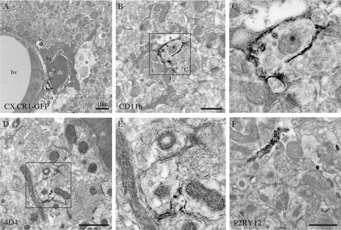
Phenotypic characterization of the dark microglia, using immunoperoxidase staining in the CA1 lacunosum‐moleculare of stressed CX3CR1 knockout mice (A–C, F), or a nontransgenic control mouse (D, E). A: Focal staining for GFP in a dark microglial cell (dc) from a CX3CR1‐GFP mouse. In contrast, normal microglia display strong and diffuse immunoreactivity for IBA1 throughout their cytoplasm. B, C: Examples of dark microglia staining for the myeloid cell marker CD11b, which forms CR3 involved in phagocytosis, strongly expressed at the plasma membrane of their processes encircling synaptic elements. D, E: Dark microglia's staining for 4D4, a recently discovered marker of homeostatic microglia, at the extremity of their ramified processes. In contrast, the dark microglia do not stain for P2RY12 (F), another marker of homeostatic microglia that is abundant in microglial processes (m). a = astrocytic process, bl = basal lamina, bv = blood vessel, s = dendritic spine, and t = axon terminals. Asterisks show the extracellular space. Scale bars = 1 μm.