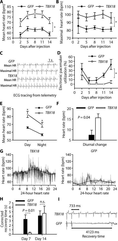
Fig. 1. TBX18 gene transfer creates a biological pacemaker.
(A and B) Mean (A) and maximal (B) HR in TBX18- and GFP-transduced animals. Data are means ± SEM (n = 5 controls, 7 TBX18). *P < 0.05 for all time points by repeated-measures analysis of variance (ANOVA). bpm, beats per minute. (C) Representative telemetry tracings of mean and maximal HR from both experimental groups. (D) Electronic pacemaker use after gene transfer. Data are means ± SEM (n = 5 controls, 7 TBX18). *P < 0.05 for all time points by repeated-measures ANOVA. (E and F) Daytime and nighttime HR (E) and diurnal changes (F) in treatment groups. Data are means ± SEM (n = 5 controls, 7 TBX18). *P < 0.05 by two-tailed t test. (G) Representative 24-hour HR trend in TBX18- and GFP-transduced animals. HR is consistently higher in daytime than in nighttime in TBX18 group. (H and I) Electrophysiological (EP) evaluation of biological pacemaker function. Corrected recovery time was measured on days 8 and 14 (H). Data are means ± SEM (n = 3 controls, 5 TBX18 at day 8; n = 5 controls, 7 TBX18 on day 14). P values were determined by two-tailed t test. n.s., not significant. Representative recovery time tracings were taken on day 8 in TBX18 and GFP animals (I).

