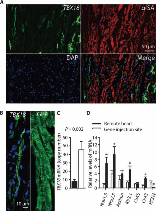
Fig. 4. TBX18 converts ventricular myocytes to iSAN cells in situ.
(A) Confocal microscopy image showing focal tissue expression of TBX18 (green fluorescence). Cardiomyocyte sarcomeres were stained with α-sarcomeric actinin (α-SA); 4′,6-diamidino-2-phenylindole (DAPI) indicates nuclei. Scale bar, 50 μm. (B) TBX18-transduced ventricular myocytes are smaller in size and spindle-shaped compared with the morphology of GFP-transduced cardiomyocyte cells. Images are representative of the injection site. (C) TBX18 mRNA expression at the gene injection site and in the left ventricular free wall (remote site). Data are means ± SEM (TBX18, n = 3, one sample per site for each animal). P value was determined by two-tailed t test. (D) Relative expression (compared to the housekeeping gene ACTA1) of iSAN-associated mRNA at gene injection site compared to the left ventricular free wall (remote site). Data are means ± SEM (TBX18, n = 6, one sample per site for each animal). *P < 0.05, determined by two-tailed t test.

