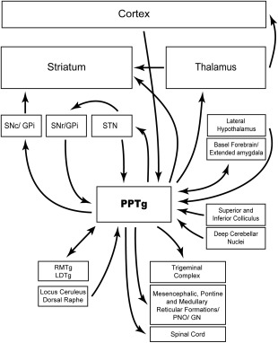
Figure 1.

Schematic illustration of pedunculopontine connectivity. Distinct functional types of pedunculopontine subpopulations innervate basal ganglia and in turn basal ganglia structures project back to different neuronal populations in the pedunculopontine. It is important to note that projections form the pedunculopontine to the structures illustrated here are not wholly independent: cholinergic and noncholinergic neurons from topographically distributed populations send collaterals to several structures (eg, to thalamus and basal ganglia). Likewise, descending collaterals of ascending axons contribute to a dense innervation of structures in the lower brainstem, pons, medulla, and spinal cord.125
