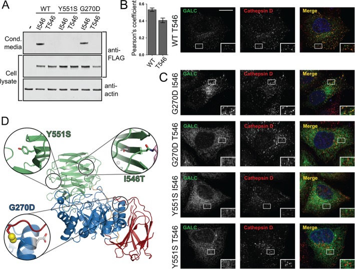
Figure 7.

Polymorphic background alters the trafficking of some missense mutations. A) HEK293T cells were transiently transfected with WT GALC (I546), T546, Y551S‐I546, Y551S‐T546, G270D‐I546, G270D‐T546. Conditioned media was harvested at 72 h and cells lysed in 1% SDS followed by SDS‐PAGE and immunoblotting. Secreted and total GALC expression was detected using western blot against the FLAG epitope tag and loading analysed by blotting against actin. B) Representative confocal microscopy images of HeLa cells transiently transfected with WT GALC containing the T546 polymorphism. Cells were plated onto glass coverslips, fixed and immunostained for GALC (green), the lysosomal marker cathepsin D (red). Nuclei were stained with DNA‐binding dye, DAPI (blue). Scale bar 10 µm. To quantify colocalization Pearson's correlation coefficients were calculated for cathepsin D. Mean ± SEM for at least 20 individual cells from ≥3 independent experiments are shown. C) Equivalent confocal microscopy images as in panel B for Y551S‐I546, Y551S‐T546, G270D‐I546 and G270D‐T546. D) The positions of I546T, G270D and Y551S are highlighted on the structure of GALC (PDB ID: 3ZR5). The structure is coloured as in Figure 5. For each variant, the zoomed view (inset) shows the relevant side chain as sticks and the surrounding region of the structure that would be affected by the mutation. The single disulphide bond (yellow) present in GALC is illustrated as spheres.
