Figure 1.

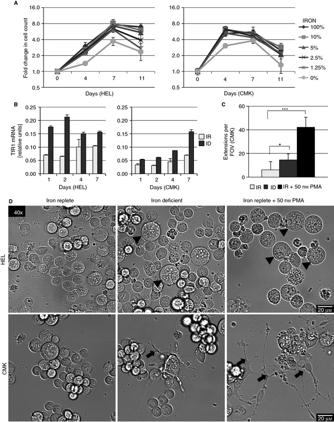
Iron deficiency (ID) leads to megakaryocytic morphological changes in HEL and in CMK. (A) Proliferation of HEL and CMK cells on culture in 100% (iron replete [IR]), 10%, 5%, 2.5%, 1.25% (ID), and 0% v/v Panserin 401 in Panserin 401S after 4, 7, and 11 days of culture. Graphs depict the fold change in cell count of propidium iodide–excluding cells measured by flow cytometry at each condition and time point as compared with day 0. (B) Relative TfR1 mRNA concentration of HEL and CMK cultured in IR or ID medium for 1, 2, 4, and 7 days (normalized to GAPDH). TfR1 is stabilized under low iron conditions. (C) Mean count of proplatelet extensions per ×40 field of view in CMK after culture for 4 days in IR, ID, and IR medium with 50 nmol L–1 PMA. (D) Representative pictures of live cell imaging (×40) of HEL and CMK cultured in IR, ID, and IR medium plus 50 nmol L–1 PMA for 4 days. Arrowheads depict large cells, which increase in ID and PMA‐treated HEL, while arrows depict formation of proplatelet‐like structures in CMK. The results from two or three independent experiments are shown. *P ≤ 0.05, ***P ≤ 0.001.
