Figure 3.

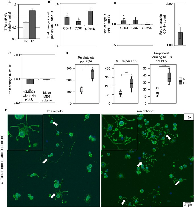
Iron deficiency (ID) potentiates megakaryocyte (MEG) differentiation in cord blood–derived hematopoietic stem cells. MEGs cultured in iron‐replete (IR) and ID media supplemented with 100 ng mL−1 TPO for 5 days. (A) Relative TfR1 mRNA concentration of MEGs cultured in IR and ID media (normalized to HPRT1). (B) Flow cytometric measurement of CD41, CD61, and CD42b after MEG culture in ID and IR media. Graphs depict the fold change in the percentage of cells expressing surface markers; the fold change in the median fluorescence intensity (MFI) of the gated positive CD41, CD61, or CD42b cells; and the fold change in the CD41+ cell count in ID‐ vs. IR‐cultured MEGs. (C) Flow cytometric measurement of ploidy by Hoechst 33342 nuclear staining. Graph depicts the fold change in the percentage of MEGs with ploidy greater than 4n and the mean MEG volume in ID compared with IR. (D) Boxplots depict proplatelet count per field of view (FOV), MEG number per FOV, and number of MEGs forming proplatelets per FOV after culture in ID and IR media. (E) Representative images (×10) of adherent MEGs forming proplatelets on fibrinogen‐coated coverslips after 5 days’ culture in IR and ID media. Arrows indicate proplatelet extensions. Insets show zoomed‐in proplatelet structures. The results from three to five independent experiments are shown. *P ≤ 0.05, **P ≤ 0.01, ***P ≤ 0.001.
