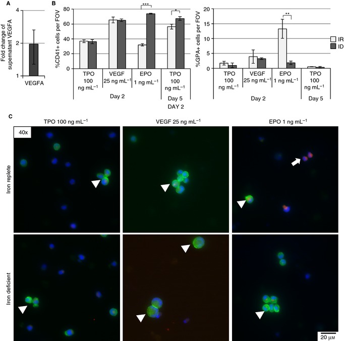
Figure 6.

VEGFA increases megakaryocyte (MEG) concentration in cord blood–derived hematopoietic stem cells. (A) Measurement of VEGFA concentration in MEG culture supernatants after 5 days of iron deficiency (ID) by immunoassay. Graphs depict the fold change in the concentration of VEGFA of ID vs. iron‐replete (IR) media. (B) Mean percent of CD41+ and glycophorin A (GPA)+ cells per field of view (FOV) of stained and fixed samples after 2‐day culture in IR and ID media supplemented with 100 ng mL−1 TPO (control), 25 ng mL−1 VEGF, and 1 ng mL−1 EPO. (C) Representative images of CD41 (green, arrowheads), glycophorin A (red, arrows), and DAPI nuclear staining after culture in IR and ID media supplemented with 100 ng mL−1 TPO (control), 25 ng mL−1 VEGF, and 1 ng mL−1 EPO for 2 days. The results from two to five independent experiments are shown. *P ≤ 0.05, **P ≤ 0.01, ***P ≤ 0.001.
