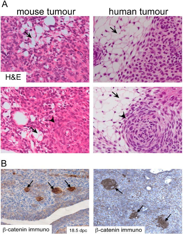
Figure 1.

Histomorphological features of mouse and human ACP. (A) Haematoxylin and eosin staining of mouse and human tumours showing the presence of microcystic changes (stellate reticulum; arrows) and whorl‐like nodular structures (cell clusters; arrowheads). (B) Immunohistochemistry with a specific anti‐β‐catenin antibody showing the presence of cell clusters with nucleo‐cytoplasmic accumulation of β‐catenin. Reproduced with permission from PNAS (Gaston‐Massuet et al., PNAS USA 2011, vol. 108, number 28, pp 11482–11487).
