
Figure 5.
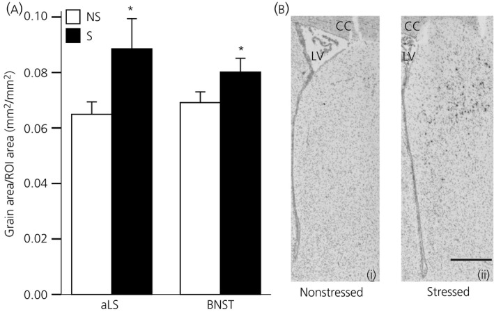
Effect of acute stress on central Avpr1a mRNA expression in prenatally stressed (PNS) females. (a) Quantification of Avpr1a mRNA expression in the anterior lateral septum (aLS) and bed nucleus of the stria terminalis (BNST) in nonstressed (ns; white bars) or stressed (restraint; black bars) PNS females. PNS females exposed to acute stress had significantly greater Avpr1a mRNA expression in both regions compared to nonstressed PNS females. *P ≤ 0.05 versus nonstress group. (b) Representative photomicrographs of Avpr1a mRNA expression in the aLS from a (i) nonstressed, and (ii) restrained PNS female. LV, lateral ventricle; cc, corpus callosum. Scale bar = 500 μm. Data are the group mean ± SEM (n = 8 rats per group). ROI, region of interest.