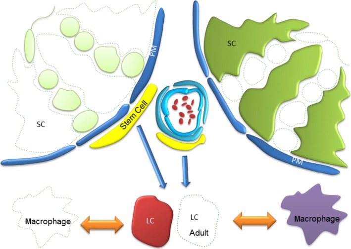
Figure 1.

Schematic representation of the testis showing ablated cell populations. To date methodologies have been employed to selectively ablate germ cells, Leydig cells, Macrophages and recently Sertoli cells. By removing a single population and asking ‘what changes’ and ‘what remains the same?’, these studies have proven instrumental in defining our fundamental understanding of cell–cell communication in the testis, assigning specific roles to individual cell types whilst ruling out others. SC = Sertoli cell; PM = Peritubular Myoid cell; LC = Leydig cell; Stem cell = Leydig stem cell.
