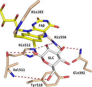
Figure 1.

Active site of Am PDH (PDB code: 4H7U) with GLC bound according to pose A (i.e. oriented for oxidation at C2). GLC coordinates from the closely related Tm POx (PDB code: 3PL8) were grafted into Am PDH after superimposing the X‐ray structures of both enzymes. Atom‐colouring scheme: carbon (beige, protein; yellow, FAD; white, ligand), nitrogen (blue) and oxygen (red). Red dashes represent important hydrogen bonds between GLC and Am PDH found during molecular dynamics simulations 17 and docking 13 in previous studies. Image generated using pymol (http://www.pymol.org/).
