
Fig. 2.
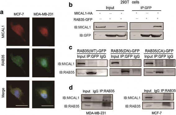
Active form of RAB35 binds to MICAL1. a Representative micrographs of MDA-MB-231 and MCF-7 cells stained for RAB35 (green) and MICAL1 expression (red) by immunofluorescence assay. Scale bar, 10 μm. b Coimmunoprecipitation experiments were performed with HEK293T cells cotransfected with HA-tagged MICAL1 and GFP-tagged RAB35. c MCF-7 cells were transfected with GFP-tagged RAB35 (WT), RAB35 (DN) or RAB35 (CA), and then immunoprecipitated with anti-GFP antibody, followed by immunoblotting analysis for RAB35 and MICAL1. d Binding of endogenous RAB35 to MICAL1 was detected in MDA-MB-231 cells and MCF-7 cells by coimmunoprecipitation experiments. n = 3 for all experiments