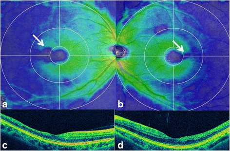
Fig. 2.

Optical coherence tomography findings for our 42-year-old Japanese patient with a 20-year history of bilateral paracentral scotoma secondary to trauma. The ganglion cell complex (GCC) shows thinning (blue; 250 μm) of the temporal macula (arrows) to the fovea (green; 300 μm) in the right (a) and left (b) eyes. Cross-sectional images also demonstrate temporal thinning of the inner retinal layers in the right (c) and left (d) eyes. The ellipsoid zone is well preserved in both eyes
