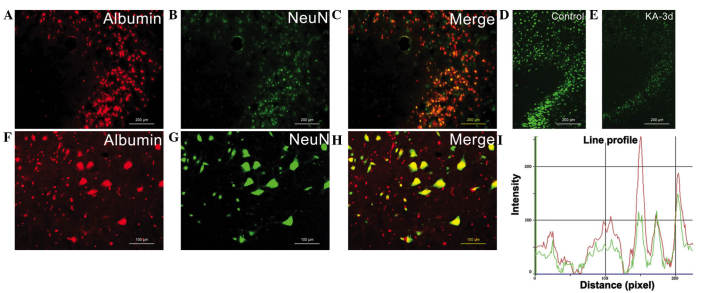
Figure 3.
Serum albumin was significantly absorbed by neurons. (A and F) Evans blue staining of kainic acid showed the extravasation of albumin. (B and G) NeuN staining of neurons. (C and H) Merged images of Evans blue and NeuN staining. NeuN expression levels were reduced in neurons following injection with (E) KA, compared with the (D) control group. (I) Line profile graph results showed an association between the immune-intensity of albumin (red) and NeuN (green) in different positions. Scale bar: (A-E), 200 µm; (F-I), 50 µm. KA, kainic acid.

