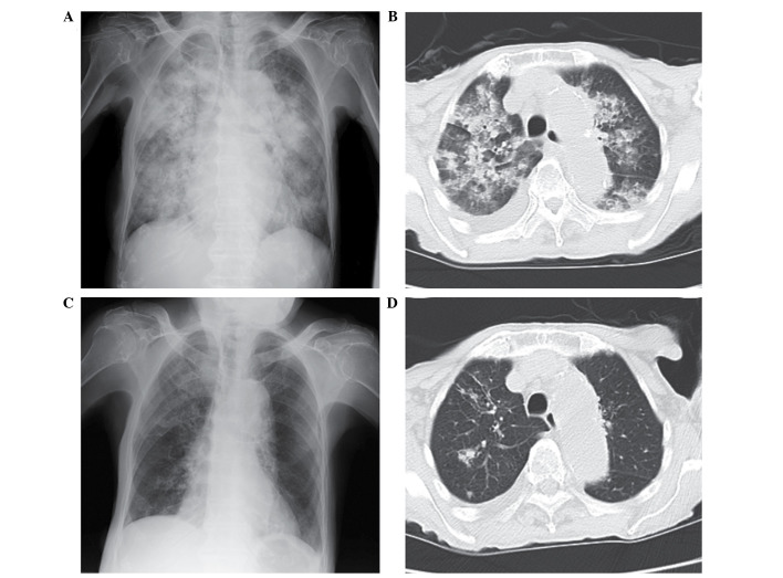
Figure 1.
(A) Chest X-ray film presenting diffuse bilateral consolidation. (B) CT scan presenting diffuse bilateral consolidation and right pleural effusion. (C) Chest X-ray film presenting near complete absence of the diffuse bilateral consolidation observed previously, with a remaining pale patchy shadow in the right upper lung field. (D) CT scan presenting nodular opacity with small satellite nodules in the right upper lobe. CT, computerised tomography.

