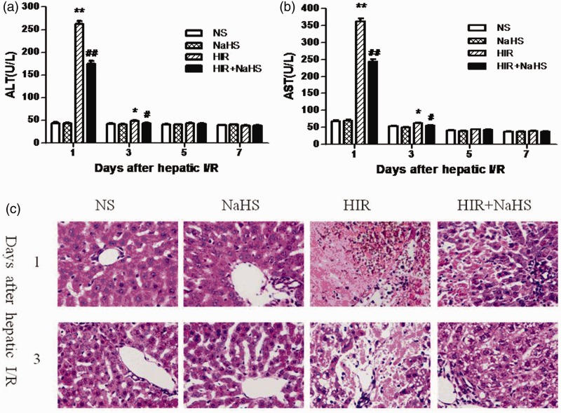
Figure 1.
NaHS attenuated hepatic I/R injury. (a) ALT level, (b) AST level, (c) representative sections of median lobe liver stained with hematoxylin–eosin were taken on the first and third day after hepatic I/R (400 × magnification). The data are expressed as the mean ± SEM. *P < 0.05 HIR versus NS, **P < 0.01 HIR versus NS, #P < 0.05 HIR + NaHS versus HIR, ##P < 0.01 HIR + NaHS versus HIR. (A color version of this figure is available in the online journal.)

