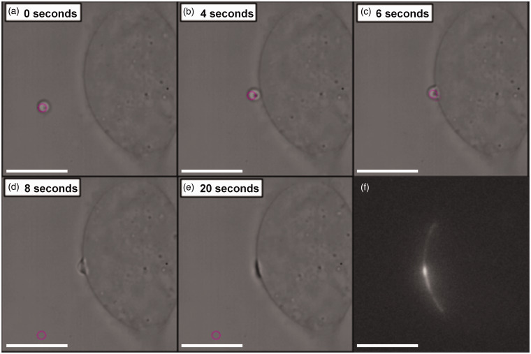
Figure 4.
Cell paintballing using coacervate microdroplets. Armstrong et al. recently demonstrated that membrane-free coacervate microdroplets can be actively loaded with biomaterial payloads of protein or nucleotides, and then delivered to the cell membrane using optical tweezers.113 (a)–(e) Time-lapse bright field microscope images showing an optical trap (pink circle) maneuvering a GFP-loaded coacervate microdroplet toward a human mesenchymal stem cell to initiate a targeted fusion event. (f) Fluorescence microscopy revealed fluorescence emission from the GFP payload present at the site of delivery

