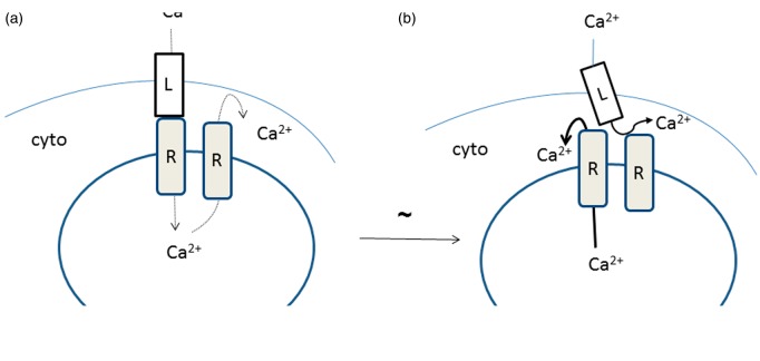
Figure 6.
Proposed mechanism for excitation–contraction coupling. In the resting state (a), a slow inward flux of Ca2+ to the SR involves both the DHPR (labelled L in the figure) and an RyR (labelled R in the figure). There is also a slow exit of Ca2+ from the SR to the cytosol (since the Ca2+ channel is inactivated during the resting state (56)) this is indicated by the thin dashed lines. In the excited state (b), movement of the DHPR exposes the Ca2+ release site of the RyR, Ca2+ rapidly flows from the SR to the cytosol. (A color version of this figure is available in the online journal.)

