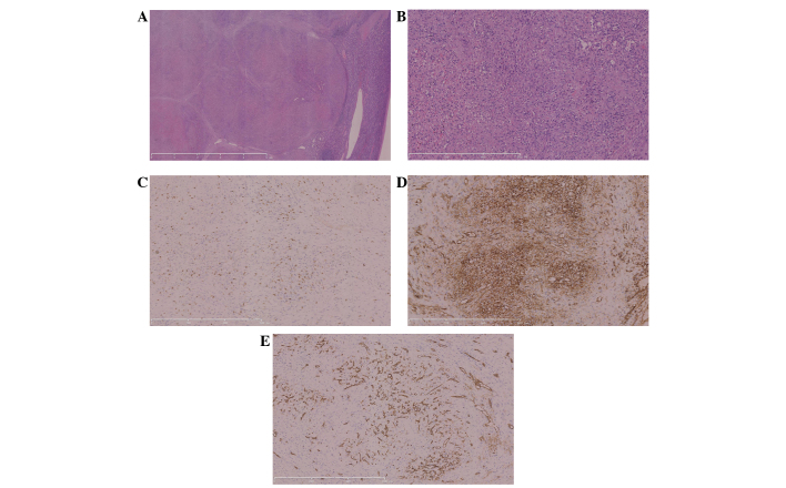
Figure 2.
Pathological examination of sclerosing angiomatoid nodular transformation. (A) Nodules were separated by fibrous or fibrosclerotic stroma (magnification, ×12.5). (B) Within the nodules, extravasated erythrocytes and hemosiderin pigment were plentiful. Immunohistochemical analysis documented positive expression of (C) CD8, (D) CD31 and (E) CD34 (magnification, ×100). CD, cluster of differentiation.

