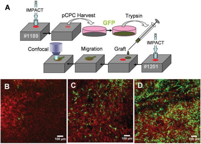
Figure 2.
Migration of grafted putative chondrogenic progenitor cells (pCPC). A, Procedure for harvesting and grafting putative chondrogenic progenitor cells. The boxes represent 2 different explants (specimen no. 1189 and specimen no. 1201). Explant no. 1189 was impacted and incubated for 5 days to allow putative chondrogenic progenitor cells to emerge. These cells were harvested and placed in monolayer culture for green fluorescent protein (GFP) transduction. Labeled cells were trypsinized, suspended in a temperature-sensitive hydrogel, and grafted onto explant no. 1201, which had been impacted a few hours earlier. B–D, The impact site was imaged by confocal microscopy at various times after grafting. Grafted GFP-labeled cells (green) can be seen against the background of host cells labeled with a red tracking stain. Exactly the same field within the impact site was imaged 2 days (B), 5 days (C), and 12 days (D) after grafting.

