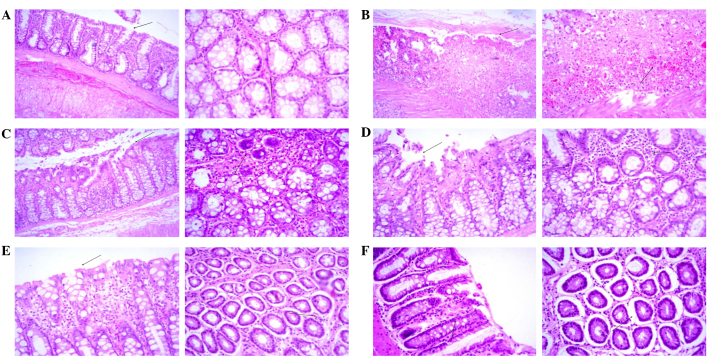
Figure 5.
Histopathological examination of colonic sections (Left panels, magnification ×10; right panels, magnification ×20). (A) Minimal surface necrosis and ulceration (arrows) with no inflammatory cell infiltration, edema or hyperemia was observed in the control group. (B) Severe surface ulceration, necrosis, edema, hyperemia and inflammatory cell infiltration (arrows) was observed in the acetic acid-treated group. (C) Moderate surface necrosis, ulceration, edema, hyperemia and inflammatory cell infiltration (arrows) was observed in the 125 mg/kg/day myrrh-treated group. (D) Mild surface necrosis, ulceration, inflammatory cell infiltration, edema and hyperemia (arrows) was observed in the 250 mg/kg/day myrrh-treated group. (E) No evidence of surface ulceration or necrosis and minimal inflammatory cell infiltration, edema and hyperemia (arrows) was observed in the 500 mg/kg/day myrrh-treated group. (F) Minimal focal surface necrosis, ulceration, edema, hyperemia and inflammatory changes (arrows) were observed in the 300 mg/kg/day mesalazine-treated group.

