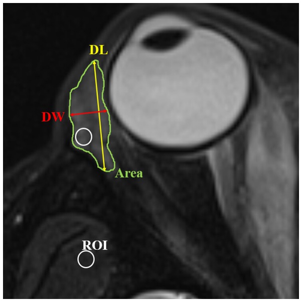
Figure 1.
Axial T2-weighted fat suppression imaging showing the methods of measurement of length, width, area, volume and SIR of the lacrimal gland. The axial image in which the lacrimal gland appeared the largest was chosen. Axial area of the lacrimal gland was obtained by manually delineating the gland border (Area). Axial length of the lacrimal gland was defined from the most anterior tip to the most posterior tip (DL). Axial width was measured from the lateral edge to the medial edge at its widest point perpendicular to the length line on the axial images (DW). Volume of lacrimal gland was obtained by a sum-of-area method. The SIR was calculated as the ratio of signal intensity of the lacrimal gland and adjacent temporalis muscle by applying a ‘hotspot’ ROI (usually 10–15% of whole cross-sectional area of lacrimal gland). The ROI placed on lacrimal gland demonstrated the portion of relatively higher signal intensity. SIR, signal intensity ratio; ROI, region of interest.

