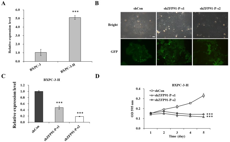
Figure 1.
Expression of ZFP91-P in pancreatic cancer cells. (A) mRNA expression levels of ZFP91-P in BXPC-3 and BXPC-3-H cells were determined by reverse transcription-quantitative polymerase chain reaction (RT-qPCR). β-actin was used as an internal control gene. ***P<0.001 vs. BXPC-3. (B) Fluorescence photomicrographs of BXPC-3-H cells infected by lentivirus. Multiplicity of infection, 20. Magnification, ×100. Scale bar, 10 µm. (C) RT-qPCR analysis of ZFP91-P mRNA expression in BXPC-3-H cells with three treatments. β-actin was used as an internal control gene. ***P<0.001 vs. shCon. (D) BXPC-3-H cell proliferation after ZFP91-P silencing, as determined by an MTT assay. Cells with three treatments including shCon, shZFP91-P-s1, and shZFP91-P-s2 groups. shCon, BXPC-3-H cells infected with control shRNA; shZFP91-P-s1, BXPC-3-H cells infected with ZFP91-P shRNA 1; shZFP91-P-s2, BXPC-3-H cells infected with ZFP91-P shRNA2. ***P<0.001 vs. shCon. Data are presented as mean ± standard error of the mean. sh, shRNA; Con, control; ZFP91-P, zinc finger protein 91 pseudogene; GFP, green fluorescent protein; OD, optical density.

