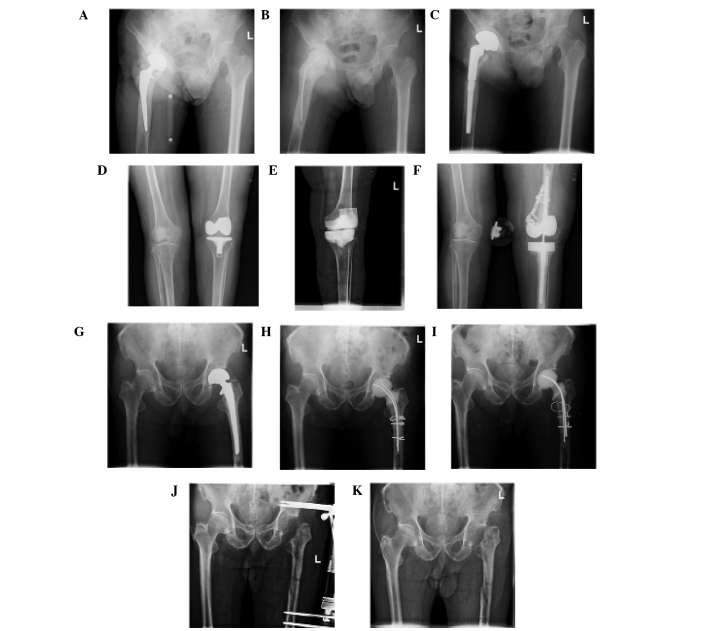
Figure 1.
Preoperative and postoperative X-rays of three cases of a fungal PJI. The X-ray images present the two-stage exchange protocol method for knee and hip joints and the method of resection arthroplasty. (A) Case 1 suffered from fungal PJI of the hip; (B) the prosthesis was removed and the cement joint spacer was implanted. (C) Following treatment with antifungal agents in the interim period, the cement spacer was removed and the prosthesis was reimplantated. (D-F) Case 6 underwent a similar therapeutic program, although the patient suffered from fungal PJI of the knee. (G-I) Case 4 was treated with the cement spacer, but then refused to receive reimplantation of the prosthesis; therefore, (J and K) resection arthroplasty was performed. PJI, prosthetic joint infection.

