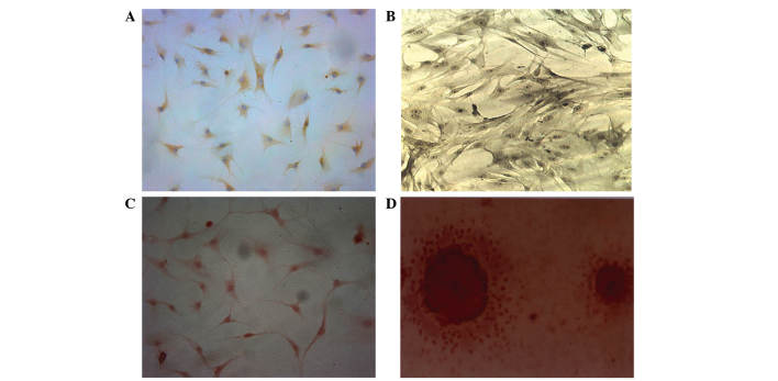
Figure 5.
Type II collagen immunohistochemical staining. (A) In the chondroblast-induced group, the cells displayed brownish yellow granules in the cytoplasm following type II collagen immunohistochemical staining (magnification, ×200). (B) In the osteoblast-induced group (magnification, ×200), gray-black granules or black deposit were observed in the (C) cytoplasm (magnification, ×200) and (D) Alizarin red staining revealed a large number of mineral nodes in the osteoblast-induced group (magnification, ×400).

