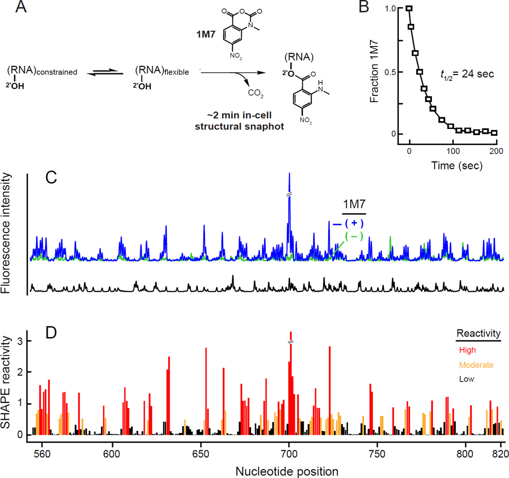
Figure 1. In vivo SHAPE of 16S ribosomal RNA.
(A) Schematic for using 1M7 to obtain ~2 min structural snapshots in vivo. (B) Pseudo-first order hydrolysis of 1M7 in Luria Broth (37 °C) at pH 7.0, corresponding to the mid-log phase of cell growth. This panel was adapted from prior work.1 (C) Electropherogram resulting from in-cell SHAPE probing of 16S rRNA. Region shown lies in the central domain. Reactions performed in the presence and absence of 1M7 are indicated by blue and green, respectively; dideoxy sequencing (ddC) trace is black. (D) Histogram of normalized SHAPE reactivities.23

