
Fig. 1.
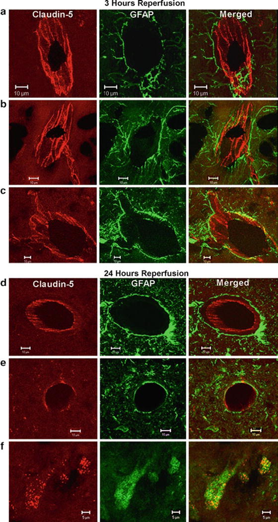
Confocal micrographs showing claudin-5 immunoreactivity in the sham-operated animals and ischemic and nonischemic hemispheres after 3 h of reperfusion (a–c). The sham-operated animals and the nonischemic side show that the claudin-5 (Cy-3, red) in blood vessels is separated from the astrocytes (GFAP-FITC, green) surrounding them (a and b). The merged images show that the claudin-5 and astrocytes are separate. In the ischemic hemisphere (c), there is fragmentation and degeneration of the claudin-5 immunoreactivity. Co-localization of claudin-5 and GFAP was seen in the ischemic hemisphere. Confocal micrographs showing claudin-5 immunoreactivity in the ischemic and nonischemic hemispheres after 24 h of reperfusion (d–f). Immunoreactivity of claudin-5 with GFAP in brain vessels in nonischemic hemisphere (d). Ischemia caused a loss of claudin-5 from the blood vessels at 24 h (e). There was co-localization of the claudin-5 in the GFAP-positive astrocytes (f). These photos were taken from penumbral areas in the caudate and lateral cortex. Scale bars indicate 10 μm in (a–e). Scale bars show 5 μm in (f) (reproduced from ref. 23).