Fig. 3.
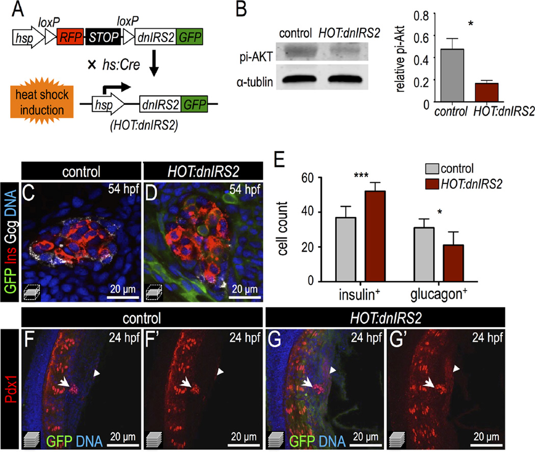
Inducible genetic blockade of insulin signaling promotes β cell formation. (A) Schematic of genetic insulin signaling blockade strategy. In double transgenic embryos, heat shock induces Cre expression and recombination of the mCherry-switch-dnIRS2-GFP transgene as well as expression of the recombined dnIRS2-GFP transgene. (B) Immunoblot of pi-Akt in Cre-negative control versus Cre-positive embryos showing that induction of dnIRS2-GFP effectively blocks insulin signaling. (C,D) Confocal planes of 54 hpf control and HOT:dnIRS2 islets stained for GFP (green), Insulin (red), and Glucagon (white). Embryos were heat shocked repeatedly, at 10, 24, 28, 32, 36, and 48 hpf; dnIRS2-GFP was induced ubiquitously, but only in Cre-positive HOT:dnIRS2 embryos. (E) Quantification of Insulin+ β cells and Glucagon+ α cells in control (gray, n = 8) and dnIRS2-expressing HOT:dnIRS2 (red, n = 6) groups. (F–G′) Merged and single channel confocal projections of 24 hpf control and HOT:dnIRS2 pancreatic endoderm stained for GFP (green) and Pdx1 (red). dnIRS2-GFP is induced ubiquitously in HOT:dnIRS2 embryos, which show expanded Pdx1 protein expression in the principal islet (arrows) and adjacent ventral endoderm (arrowheads). Student t-test was used in B and two-way ANOVA was used in E to determine significance.

