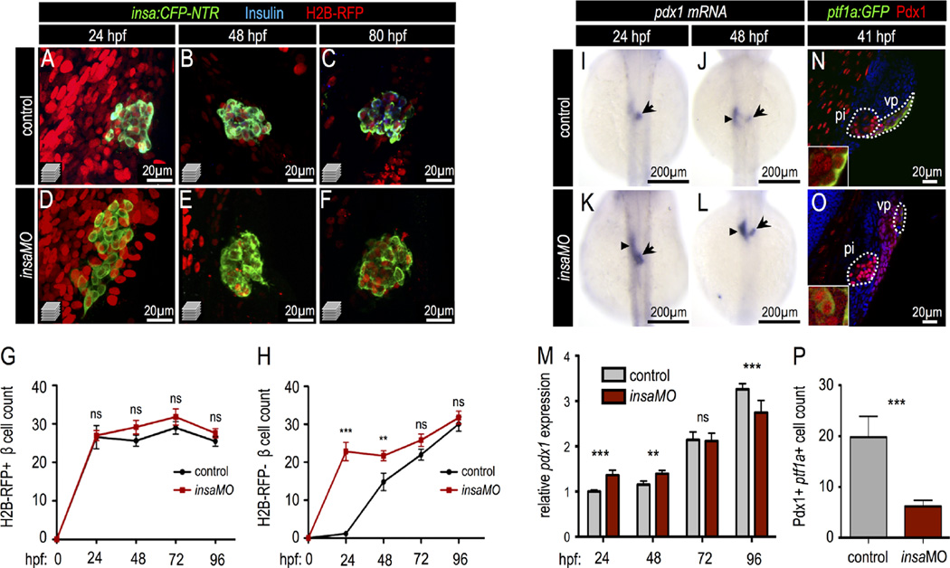
Fig. 4.
Insulin knockdown increases Pdx1 expression and drives early pancreatic progenitor differentiation into β cells. (A–F) Confocal projections of 24 hpf, 48 hpf, and 80 hpf Tg(insa:CFP-NTR) control (A–C) and insaMO-injected (D–F) islets that were co-injected with H2B-RFP mRNA to distinguish dorsal (H2B-RFP-positive DBCs) and ventral (H2B-RFP-negative VBCs) bud-derived β cells. Islets were immuno-stained for CFP (β cells, green) and insulin (blue). Insulin protein was not detectable in insaMO-injected islets. (G,H) Quantification of H2B-RFP-positive DBCs (G) and H2B-RFP-negative VBCs (H) in control (black line) and insaMO-injected (red line) islets from 24 hpf to 96 hpf (n ≥ 8 islets at each stage). VBC count is increased early at 24 and 48 hpf, but normalizes to control levels by 72 hpf. (I–L) In situ hybridization of pdx1 expression in 24 hpf and 48 hpf control (I,J) and insaMO-injected (K,L) embryos. pdx1 expression was increased in the principal islet (arrow) and adjacent endoderm (arrow heads). (M) Real time PCR to detect Pdx1 expression in both control and insaMO-injected embryos from 24 hpf to 96 hpf (n = 3). (N,O) Confocal planes of 41 hpf Tg(ptf1a:GFP) control (N) and insaMO-injected (O) endoderm stained for GFP (green) and Pdx1(red). Pdx1 expression was increased while ptf1a:GFP expression was decreased in insaMO-injected embryos. (P) Quantification of double positive Pdx1+ ptf1a+ pancreatic progenitor cells in both control (n = 6) and insaMO (n = 6) injected embryos. Abbreviations: pi, principal islet; vp, ventral pancreas. ANOVA was used in G,H, and O and Student's t-test was used in P to determine significance.

