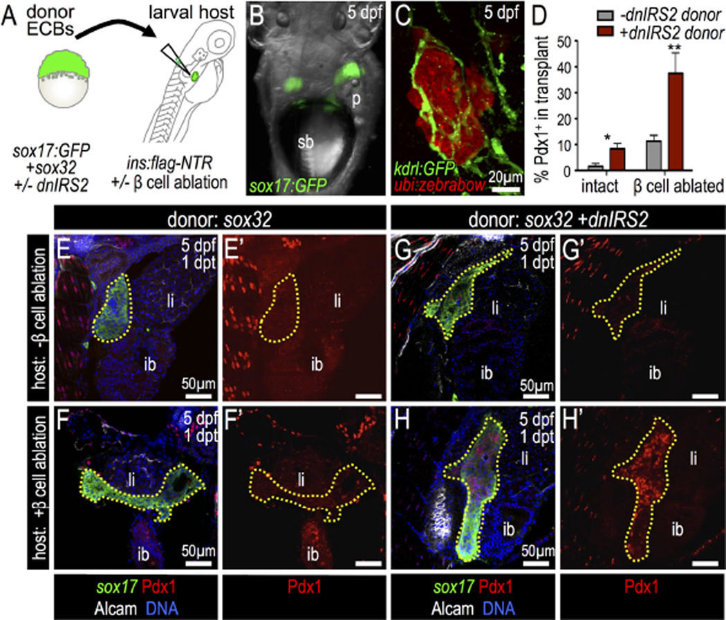
Fig. 8.
Insulin signaling blockade in endodermal committed blastomeres promotes Pdx1 expression after transplantation into β-cell ablated hosts. (A) Blastula ECB to 4 dpf Tg (insa:flag-NTR) larva cell transplantation scheme. (B) 5 dpf host shows Tg(sox17:GFP) donor cells at 1 day post transplantation (dpt). (C) Confocal projection of Tg(kdrl:GFP) (green) host with transplanted Tg(ubi:zebrabow) donor cells (red) at 1 dpt shows engraftment. (D) Percentage of transplanted ECBs that are Pdx1-positive. Chimeras were generated with the following four combinations: (1) wild type ECBs (−dnIRS2) into non-ablated host (n = 4); (2) ECBs +dnIRS2 into non-ablated host (n = 6); (3) wild type ECBs (−dnIRS2) into β ablated host (n = 6); and (4) ECBs +dnIRS2 into β ablated host (n = 8). (E–H′) Merged and single channel confocal planes of chimeras showing Tg(sox17: GFP) donor ECBs (outlined) at 1 dpt, stained for GFP (green), Pdx1 (red), Alcam (white), and DNA (blue). (E–F′) sox32 mRNA-injected blastomeres transplanted into β intact (E′ E′) and β cell-ablated hosts (F–F′). Few transplanted ECBs expressed Pdx1. (G–H′) sox32 and dnIRS2 mRNA-injected blastomeres transplanted into β cell-intact (G–G′) and β cell-ablated (H–H′) hosts. Loss of β cells increases Pdx1 expression in transplanted ECBs. Student t-test was used for statistical analysis in D. Abbreviations: sb, swim bladder; p, pancreas; ib, intestine bulb; li, liver.

