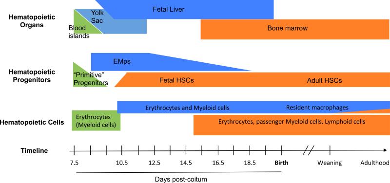
Figure 1.
Time scale of hematopoietic progenitors development in the mouse embryo.There are three successive but overlapping waves of hematopoietic progenitors during development, all of which have the potential to give rise to fetal macrophages. They can be distinguished by the hematopoietic niches exist where the progenitors expand and differentiate during development (upper panel), the yolk sac (primitive progenitors in green and EMP in blue), the fetal liver (EMP and HSC), and finally the bone marrow (HSC, orange). While primitive progenitors are only described in a small time window (middle panel), definitive progenitors (including EMPs and HSCs) co-exist during most fetal development in the fetal liver. Only HSC-derived hematopoiesis then shifts to the bone marrow niche. The three waves of progenitor can also be distinguished by their differentiation potential in vivo (lower panel). Primitive progenitors are restricted to the erythroid or the myeloid lineages, while EMP have both erythroid and myeloid potential. EMP-derived hematopoiesis gives rise to erythrocytes, macrophages, monocytes, granulocytes and mast cells and is sufficient to support survival of embryos lacking HSCs until birth. EMP-derived macrophages persist after birth in most tissues as resident macrophages, with the exception of the intestine lamina propria. HSCs are distinguished from other progenitors by their capacity to perform long-term repopulation of all leukocytes lineages when transplanted into a conventional irradiated recipient mouse.

