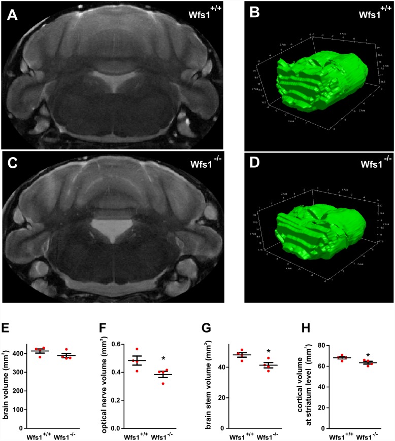
Fig 7. WFS1 deficiency is associated with reduced volume of the optic nerve, brain stem, and cortex at the level of the striatum.
(A–D) Brains, within skulls, were scanned ex vivo using a 94/20 Bruker BioSpec MRI. Representative single coronal slice from the imaging sequence (A, C) and a 3D reconstruction of the brain stem (B, D). The outer skull, tissue, and surrounding medium have been removed for clarity. (E–H) Volumetric analysis of brains from Wfs +/+ and -/- male mice: whole brain (E), optic nerve (F), brain stem (G), and cortical volumes at the level of striatum (H). *p < 0.05 and **p < 0.01 compared with Wfs +/+, groups (n = 4 animals in each group).

