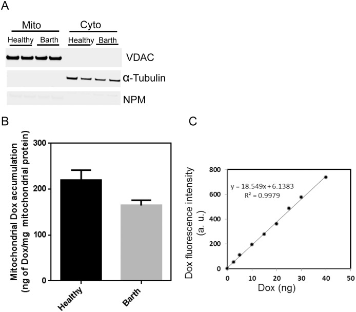
Fig 2. Mitochondrial accumulation of Dox in healthy and Barth lymphocytes.
(A) Representative western blot showing the purity of the mitochondrial fractions. Protein lysates from cell fractionation were loaded in duplicate for healthy and Barth lymphocytes. (B) Quantitative determination of mitochondrial Dox by fluorescence method (λex = 478 nm λem = 594 nm) in healthy and Barth cells. Cells were incubated with 1μM Dox for 24 h, washed with PBS, then incubated in fresh media without Dox for 2 h, then cells were harvested and subjected to isolate the mitochondrial fractionation for Dox measurement. (C) Standard curve generated from the known concentrations of Dox solutions that was used to quantify mitochondrial Dox.

