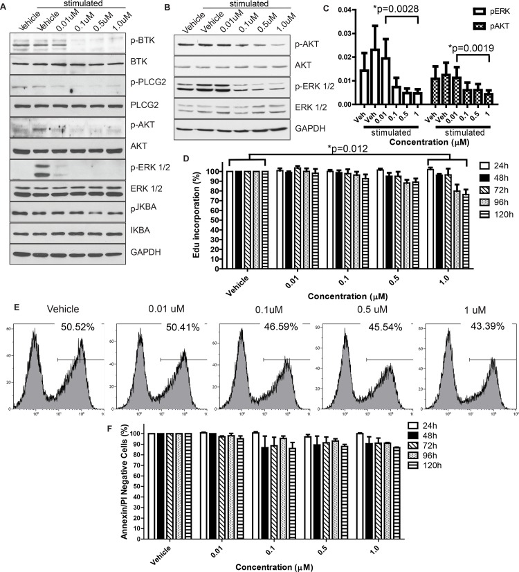
Fig 1. Effects of acalabrutinib inhibitors on canine lymphoma cells.
Dose-dependent inhibition of BTK autophosphorylation, in addition to downstream targets, was observed via immunoblot at drug concentrations as low as 0.01μM following 1 hour of treatment with acalabrutinib in the canine B-cell lymphoma CLBL1 cell line (representative of 3 repetitions) (A) and primary canine lymphoma cells treated ex vivo (representative of 4 patients, separate from the clinical study population) (B). C. Densitometry quantification of the western blots from B. Bands of phospho-proteins are normalized to respective total proteins and loading control. There was a significant dose-dependent decrease in phosphorylation for p-ERK (P = 0.0028) and p-AKT (p = 0.0019). D. Dose-dependent reductions in cell proliferation following daily treatment with acalabrutinib in the canine CLBL1 B-cell lymphoma cell line. Results are the mean of 5 independent experiments. Raw data were log transformed to reduce variance and skewness. Linear mixed effects models were applied to apoptosis data and the log-transformed proliferation data to account for the correlation of the observations from the same batch. p = 0.012 E. Representative histograms showing a dose-dependent reduction in Edu incorporation from a single day at the 72 hour timepoint. F. Dose-dependent trend toward reductions in cell viability in CLBL1. Results are the mean of 3 independent experiments. Effects not statistically significant in a linear mixed effects model.

